Heart Electrophysiology


Requires a Wolfram Notebook System
Interact on desktop, mobile and cloud with the free Wolfram Player or other Wolfram Language products.
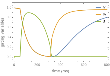
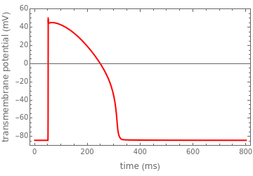
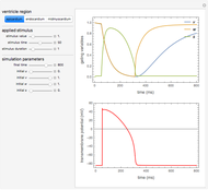
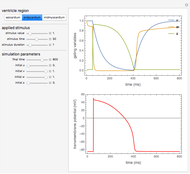
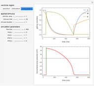
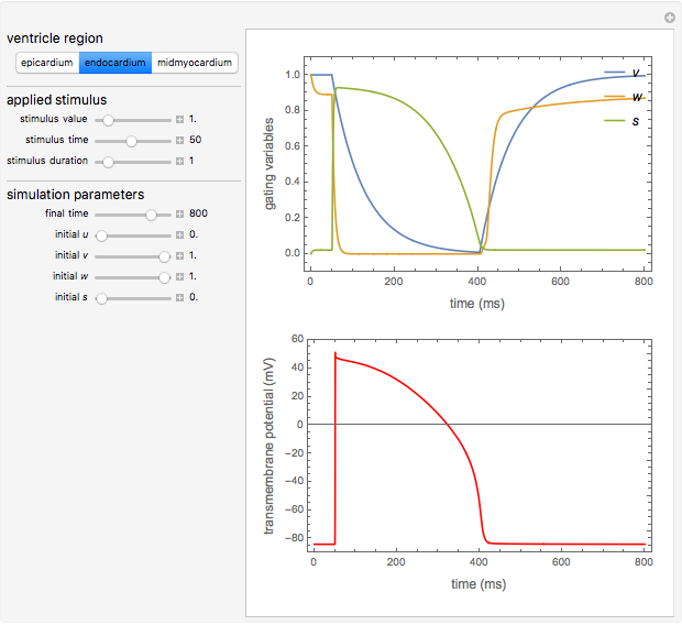
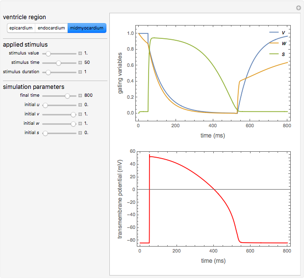
The heart wall is composed of three layers: the epicardium, myocardium and endocardium. This Demonstration shows transmembrane potentials and gating variables during a heartbeat for cardiac cells located in each of these layers. All time values are measured in milliseconds (ms).
Contributed by: Mikel Landajuela (January 2018)
Open content licensed under CC BY-NC-SA
Snapshots

Details
We consider the minimal model proposed in [1] to reproduce experimentally measured characteristics of human ventricular action potential.
Reference
[1] A. Bueno-Orovio, E. M. Cherry and F. H. Fenton, "Minimal Model for Human Ventricular Action Potentials in Tissue," Journal of Theoretical Biology, 253(3), 2008 pp. 544–560. doi:10.1016/j.jtbi.2008.03.029.
Permanent Citation
"Heart Electrophysiology"
http://demonstrations.wolfram.com/HeartElectrophysiology/
Wolfram Demonstrations Project
Published: January 16 2018
 Windkessel Model for Hemodynamics in Arterial Systems
Mikel Landajuela Factors Affecting Blood Flow
Juliet Foote and Mihir Surapaneni Transdermal Drug Delivery by Diffusion
Benjamin Pinsky and Elizabeth Le Diffusion of Oxygen through the Placenta
Colleen Tacubao, Patrick Chen and Hazel Wong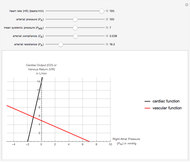 Steady-State Operation of the Cardiovascular System
Christopher B. Arena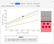 Effect of Altitude on Alveolar Oxygen Pressure
Natalie Gurevich and Kaitlyn Kuder
Ningxian "Nina" Fan and Daphne Cay Cantuba Simulating Gas Exchange in a Model of Pulmonary Fibrosis
Alexandra Peirce, Stephen Dowker and Sonia Parikh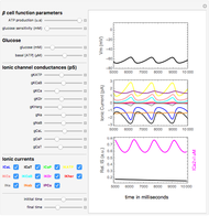 Model of Electrical Activity in Human Pancreatic Beta Cells
Gerardo J. Felix-Martinez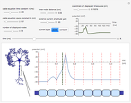 Action Potential Propagation along Myelinated Axons
Oliver K. Ernst