Gradient Echo Pulse Sequence in Magnetic Resonance Imaging

Requires a Wolfram Notebook System
Interact on desktop, mobile and cloud with the free Wolfram Player or other Wolfram Language products.
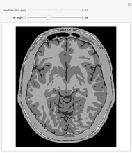
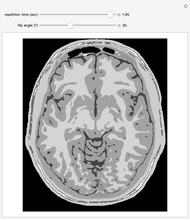
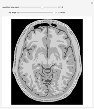
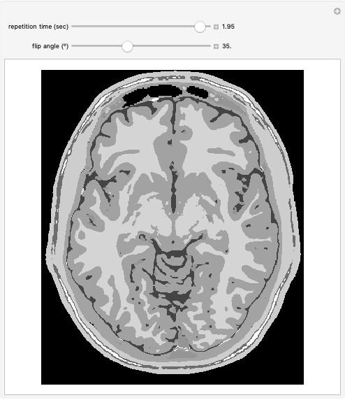
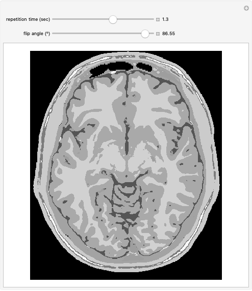
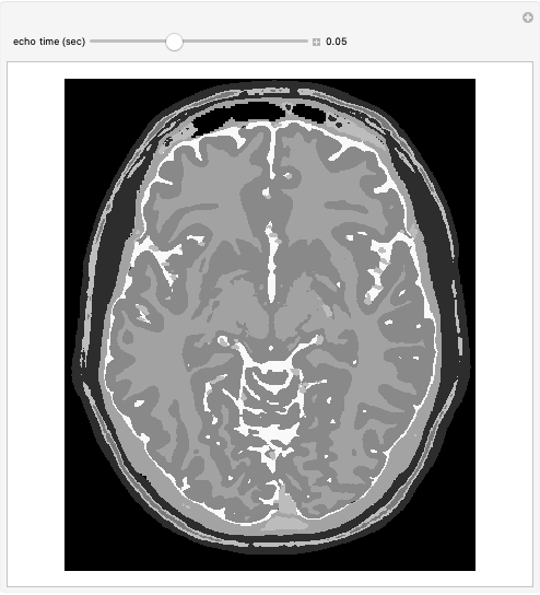
This Demonstration shows a spoiled gradient echo pulse sequence from a magnetic resonance imaging (MRI). The "repetition time" (in seconds) and "flip angle" are parameters that can be controlled on an MRI machine to yield scans with different contrast between tissues. This Demonstration examines an image of the brain with the tissues: cerebrospinal fluid, gray matter, white matter, fat, muscle, skin, dura, marrow and vessels at a magnetic field of 3.0 T.
Contributed by: Yiling Hu (August 2022)
Open content licensed under CC BY-NC-SA
Snapshots

Details
References
[1] L. Collins. "Colin 27 Average Brain 2008." McConnell Brain Imaging Centre, Montreal Neurological Institute, McGill University. (Sep 7, 2021) nist.mni.mcgill.ca/colin-27-average-brain-2008.
[2] "Tissue Properties: Relaxation Times." IT'IS Foundation. (Sep 7, 2021) itis.swiss/virtual-population/tissue-properties/database/relaxation-times.
[3] J. P. Hornak, The Basics of MRI, Chapter 10. (Sep 7, 2021) www.cis.rit.edu/htbooks/mri/chap-10/chap-10.htm.
[4] D. W. McRobbie, E. A. Moore, M. J. Graves and M. R. Prince, MRI from Picture to Proton, New York: Cambridge University Press, 2007.
Permanent Citation
 Spin Echo Pulse Sequence in Magnetic Resonance Imaging
Yiling Hu Magnetic Resonance Imaging (MRI)
Yuncong Ma Bit Response of a PR4 Encoded Magnetic Medium
Charles Masenas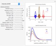 Optical Properties of Biological Tissue in the THz Radiation Regime
Muhamad Hamdi and Yusof Munajat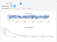 Processing a Neurological Multiunit-Activity Signal
Subashini Lakshmanan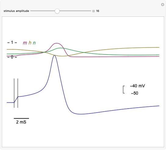 Hodgkin-Huxley Action Potential Model
Shimon Marom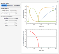 Heart Electrophysiology
Mikel Landajuela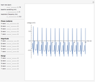 Synthetic ECG
Xiaopeng Zhao Single Signals in Nuclear Magnetic Resonance
Chengchen Guo and Jeffery L. Yarger Multiple Signals in Nuclear Magnetic Resonance
Chengchen Guo and Jeffery L. Yarger
